
The word anatomy is derived from two Greek words ana (means as under) and tomy (means to cut). It is the study of the internal structure of the body. Plant anatomy deals with the ontogeny, structurean distribution of various
types of tissues in roots, stems, leaves, flowers and fruits.
Plant anatomy is an important basic science. It is helpful in understanding the complexity of plant structure, adaptations, roles of various tissues in physiology and determination of phylogeny.Anatomical features of the vegetative and flowering organs have been successfully employed in solving taxonomic problems. Anatomical
characters of fragmentary material have proved identity of criminals) and pharmacognosy useful in forensic science (establishing correct a determining correct identity of plants of medicinal uses).

Table of Contents
TISSUES
The basic structural unit of multicellular organisms is the cell. In higher organisms, the cells are organised into tissue, tissues into tissue systems and systems into organs. Tissue can be defined as a group of cells which are essentially similar in structure and function and have a common origin. In the beginning most of the tiSsues occur
as undifferentiated group of embryonic cells capable of continuous divisions. These are known as produces meristematic tissues.undifferentiated tissue which may at a later stage, differentiate and acquire the mature state. Such called mature or permanent. tissues are Permanent tissues form Characteristic internal organization of plant organs. Permanent tissue undergo irreversible differentiation, such as sieve elements which have no nucleus and dead cells (e.g., sclerenchyma, tracheids and vessels). Thus on the basis of the ability of cells to divide, tissues can be classified into two major groups:
(1) Meristematic tissues and
(2) Permanent tissues
MERISTEMATIC TISSUES
The fertilised egg that forms an embryo consists of dividing cells. However, at later stages only a few of these cells continue to divide and help in growth. The rest of the cells differentiate and perform specific functions. Thus meristem Is in fact a part of the embryonic tissue that continues to remain embryonic- retains ability to divide.
During this period, the meristematic tissue produces leaves, branches, flowers, etc. Such a localised group, composed of young cells with an ability of continuous divisions, is known as meristematic tissue and the region as meristem.
Characteristics of Meristematic Cells
The following are the important characters of meristematic cells:
(1) Meristematic cells are thin walled,
(2) They are compactly arranged without intercellular spaces.
(3) They are densely cytoplasmic with a prominent nucleus.
(4) The vacuoles are either small or absent.
(5) Endoplasmic reticulum and mitochondria are not fully developed.
(6) They possess plastids in protoplastid stage.
(7) Metabolically, these are the most active cells.
(8) These are immature and undifferentiated which remain embryonic in cells character, i.e., retain the ability to divide.
Classification of Meristems
Meristems are classified on the basis of growth, plane of division, development, position and function of the constituting cells.
[I] Meristems based on growth
- Promeristem (Primordial meristem, embryo meristem or urmeristem). Promeristem is situated at the tips of roots and shoots. It is the first representing formed meristematic region represc embryonic stage for other developing merisems.
The derivatives give rise to all types of primary tissues.
[II] Meristems based on plane of division
Based on the plane of division, following three types of meristems have been recognised.
- Mass meristem. The cells of this meristem divide in almost all the planes producing structures with irregular shapes (e.g., endosperm, etc.).
- Plate meristem. The cells of plate meristem divide anticlinally to form plate-like Structures and form uniseriate epidermis and multiseriate blade of the leaf.
- Rib meristem. The cells of this meristem divide at right angles to the longitudinal axis
[II] Meristems based on origin
Based on the origin, meristems are of following two types.
- Primary meristem. The cells of this meristem develop directly from the embryonic cells. The cells divide and derivatives form permanent tissues. It occurs at the apices of root and shoot (apical meristems), at the base of leaves (intercalary meristem) and intrafascicular cambium in thevascular bundles (lateral meristem).
- Secondary meristem. The permanent tissue formed by the primary meristem may later become meristematic. This is called secondary meristem. For example, pericycle is a permanent tissue formed by the primary meristem. It becomes meristematic later to form a secondary meristem
the cork cambium that produces Scondary cortex. Interfascicular cambium is also a secondary meristem being formed later between the two vascular bundles.
[IV] Meristems based on position
Based on their position in the plant body, meristems can be classified into following three categories.
- Apical meristems. These are situated at the tips of the root and shoot. It causes increase in height. The examples include promeristem and primary meristem (root and shoot apices).
- Lateral meristem. The vascular cambium and cork cambium are the two examples of lateral meristem, being placed along the side of the central longitudinal axis of the plant. Vascular cambium increases the girth of the plant by producing secondary vascular tissues throughout
the period of plant life. The cork cambium or phellogen which develops later by redifferentiation of the permanen Which tissues (mostly pericycle) is a (secondary) latera meristem. . - Inter calarymeristem Intercalary meristem is located away from the apical meristem between the two differentiated region s. It is the residual part of the apical meristem that retains its meristematic activity. The examples and internodes of many leaves the include monocotyledons, the flowering Scapes and pedicels of some species, gynophore of Arachis
(ground nut), below the node in Mentha, at the base of leaf in pines, etc. In fact grass stem elongates due to the activity of intercalary meristem.
[V] Meristems based on functions
Following three types of meristems are recognised on the basis of function.
- Protoderm. This is the outermost layer that forms the epidermis.
- Procambium. The cells of this region are vertically elongated. These produce primary vascular tissues.
- Ground meristem. The cells of this region are large, thin walled and isodiametric. These produce hypodermis, cortex, endodermis, pericycie medullary rays and pith.Such an organization is mainly shown by the developed embryo and apices of shoot and rool.
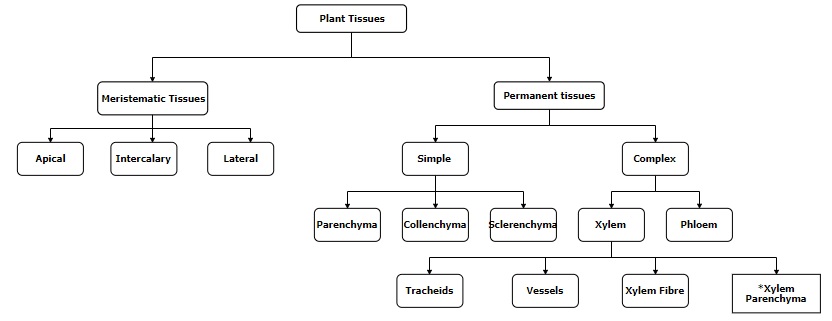
PERMANENT TISSUES
Permanent tissue may be defined as a group of cells which have lost the capacity of division Permanent tissues developing from primary meristematic tissue (i.e., promeristem) are called primary permanent tissues (e.g., parenchyma,collenchyma, etc). Similarly, permanent tissuesdeveloping from secondary meristematic tissues
(i.e., cork cambium) are known as secondary permanent tissues (e.g., cork, secondary xylem and secondary phloem).Permanent tissues can be divided into two groups:
(1) Simple tissues and
(2) Complex tissues.
Simple Tissues
Tissues compose of only one type of cells are called simple tissues.These in occur homogeneous groups. Simple tissues include:
(1) Parenchyma,
(2) Collenchyma and
(3) Sclerenchyma.
Parenchyma
- Origin. The parenchyma present in cortex, pith, mesophyll and some parts of flower is mostly producedby the ground meristeme. The parenchyma, found in primary and secondary vascular tissues, originates from procambium and respectively. Phelloderm
vascular cambium, (secondary cortex) formed by phellogen (cork cambium), also consists of parenchyma.
Characteristics of parenchyma.
Following important are the parenchyma.
(1) Parenchyma are thin walled living cells.
(2) Their cell wall is made of cellulose,Some pectin.and
hemicellulose parenchymatous cells, as those of endosperm, secondary wall of possess a also may cellulose.
(3)They are more or less spherical but may also be polyhedral having as many called sides (hence fourteen as
tetrakaidecahedron). Besides this, elongated and lobed parenchyma are also found.
(4) Young parenchymatous cells are loosely arranged and have intercellular spaces.However, mature cells being tightly packed,lack intercellular spaces.
(5) Photosynthetic parenchymatous cells have Non-photosynthetic chloroplasts. parenchyma cells possessleucoplasts. Storage parenchyma shows starch grains,inulin crystals and raphides.
Specialized types of parenchyma.
Following are the various types of parenchyma: assimilatory
(a) Chlorenchyma or parenchyma. These cells abundant possess chloroplasts. Chlorenchyma is found in leaves and cortex of young stems.
(b) Palisade parenchyma. These are elongated chlorenchymatous cells mainly found in leaves
(c) Spongy parenchyma This is a chlorenchyma of irregular shapes and sizes, with numerous intercellular spaces. It is found in the leaves.
(d) Aerenchyma. A parenchyma with well developed intercellular spaces which form a connected system throughout the plant is known as aerenchyma. It is common in submergcd hydrophytes.
(e) Prosenchyma. These are slightly thick walled elongated parenchymatous cells. They provide mechanical support to the organ in which they occur.
(f) Xylem and phloem parenchyma. These are the parenchymatous cells associated with xylem and phloem, respectively. These cells also originate from the same meristem which gives rise to xylem and phloem. These cells are thin or thick walled and are the only living cells in xylem tissue.Xylem and phloem parenchyma are meant for
storage of starch and fatty food. Sometimes they also contain tannins.
(g) Idioblast. It is a special cell which differs markedly in form, size or contents from other cells in the same tissue. Idioblasts mostly store ergastic substanes. e.g., tannin cells, cells filled with oil, crystals, raphides, etc., and mostly storeergastic substances.
- Distribution. Parenchyma is mostly found in the cortex and pith of roót and stem, mesophyl of leaves, fleshy parts of succulent fruits,endosperm of seeds, etc. It also occurs in the xylem, phloem and medullary rays.
Functions.
The following are the major functions of parenchyma.
(1) Parenchyma always retains its meristematic character and is, therefore, a potential meristematic tissue. Parenchyma becomes meristematic during wound healing, formatic of adventitious roots, grafting, etc.
(2) Photosynthesis, respiration,storage,secretion,assimilation etc., are some of the important processes which occur in parenchymatous cells.
(3) Aerenchyma helps hydrophytes to float and exhange gases due to air present in the large intercellular spaces ormed by them,
(4) In succulent xerophy parenchyma have mucilage. This helps the plant in storing water absorbed by mucilage of the cells
(5) These cells help in the lateral conduction of water and food material.
(6) Prosenchyma provides rigidity and strength to the organs in which it occurs.
CollenchymaOrigin.
Simple collenchyma originates from the ground meristem. However, some specially formed collenchymatous cells develop from procambium.
Characteristics of collenchyma
Following are the major characteristics of collenchyma
(1) It is a mechanical tissue that consists of living cells. It retains protoplasm even at maturity. The cells are polygonal, spherical or elongated with pointed or rounded ends.
(2) The cell wall is generally unevenly thickened due to deposition of pectin and hemicelluloses.
(3) Collenchyma may or may not leave intercellular spaces.
4) These may contain chloroplasts and canry out photosynthesis.
Types of collenchyma.
Following three types of collenchyma are recognised on the basis of thickening or deposition:
(a) Angular collenchyma. This is the most common type of collenchyma. The thickening occurs predominantly at the corners or angles of the cells, e.g., stems of Cucurbita, Datura, etc. The intercellular spaces may thus be altogether absert.
(b) Lamellar or plate collenchyma. In this type, thickening is deposited more heavily on the tangential walls than on the radial walls of the cells. The deposition thus appears stratified or lamellated; e.g., stems of Sambucus, Rhamnus, etc.
(c) Lacunar or tubular collenchyma. In these cells thickening is deposited primarily on the wails around the intercellular spaces, e.g.. aerial roots of Monstera and stem of Calotropis.
Distribution. Collenchyma is generally epidermis in herbaceous below the present dicotyledonous stems. It occurs either in isolated patches or in the fornm of a continuous cylinder.It is found in the petiole and lamina of the leaves. in leaves it also forms the bundle sheath.
Functions. Major functions of collenchyma are as follows:
(1) Collenchyma being flexible, provides tensile strength to various plant organs. Hence, it is primarily mechanical in function (in climbers).
(2)It has a considerable degree of plasticity and thus functions as supporting tissue.
(3)Chloroplast containing collenchyma cells also perform photosynthesis.
(4) It also serves as a storage tissue,
Sclerenchyma
- Origin. Sclerenchyma develops either from the procambium or secondarily from parenchyma due to secondary wall deposition.
- Characteristics of sclerenchyma
Following are the characteristics of sclerenchyma.
(1) The cells are long or short, narrow, thick walled and lignified.
(2) These are dead cells and do not perform any metabolic function.
(3) Theyshow different types of lignin depositions and also have pits.
Types of sclerenchyma.
Sclerenchyma may be sub-divided into sclereids and sclerenchymatous fibres
(a) Sclereids. Short or irregular sclerenchyma cells are called sclereids or sclerotic cells. Their walls are very thick, irregular and the lumen is very narrow. The walls show simple pits.
On the basis of their shape, sclereids are classified into many categories:
(i) Brachysclereids. These are isodiametric in shape and are also called stone cells, sclerotic cells, or grit cells. They occur in the pulp of some fruits; e.g., pear.
(ii) Macrosclereids. These are elongated rod- like or columnar and occur in the seed coats of pulses.
(iii) Osteosclereids. These are bone shaped, usually enlarged at the ends. They occur in the seed coats of many dicotyledons.
(iv) Astrosclereids. These are more or less star-shaped and are found in the leaves and stems of many dicotyledons.
(v) Trichosclereids. These are elongated and more or less trichome or hair-like. These are found in aerial roots of Monstera sp.
(b) Sclerenchymatous fibres.
These sclerenchymatous cells are generally many times longer than broad. Most fibres are elongated and their ends are pointed. These have a narrow lumen and thick secondary walls. The walls may possess simple, oblique or bordered pits.
According to their position in the plant body,fibres are classified into two groups xylary or xylem fibres and extraxylary fibres.Xylem fibres, are associated with the xylem.Extraxylary fibres occur in the tissues other than xylem, i.e., in the phloem or in the cortex around vascular bundles, etc.
In monocotyledons, fibres may form bundle form bundle sheath in the leaves, and are known as leaf fibres or hard fibres.
Some of the economically important fibres and their sources are as follows:
- Phloem fibres or bast fibres. Important sources of phloem fibres are:
(1)FlaX (Linum usitatissimum),
(ii) jute (Corchorus capsularis),
(iii) madras hemp (Hibiscus cannabinus),
(iv) hemp (Cannabis sativa),
(v) sunn hemnp(Crotalaria juncea),
(vi) ramie (Boehmeria nivea),etc. - Leaf fibres. Important sources of these
fibres are:
(i) manila hemp (Musa textilis),
(ii) sisal hemp (Agave sisalina), etc.
Individual fibres may attain a considerable length, e.g., 1-10 cm in hemp and up to 55 cm ramie. These fibres are separated by a decomposition process called retting. This helps in separating the fibres from other plant material. - Distribution. Sclerenchyma is widely distributed in various plant organs in groups or in layers. Besides, these cells also occur singly. Functions. Following are the major
functions of sclerenchyma:
(1) The most important function of sclerenchyma is to provide mechanical strength to the plant.
(2) These cells, being dead, do not require water.Hence, abundance of these cells in xerophytes water their reduces requirement.
(3) In many fruits sclereids form the endocarp.
(4) Distribution of sclereids in the fruit wall determines the dehiscence of fruit.
Complex Tissues
Tissues composed of two or more types of cells contributing to a common function are called complex tissues. Xylem and phloem which together form the vascular bundle are complex tissues.
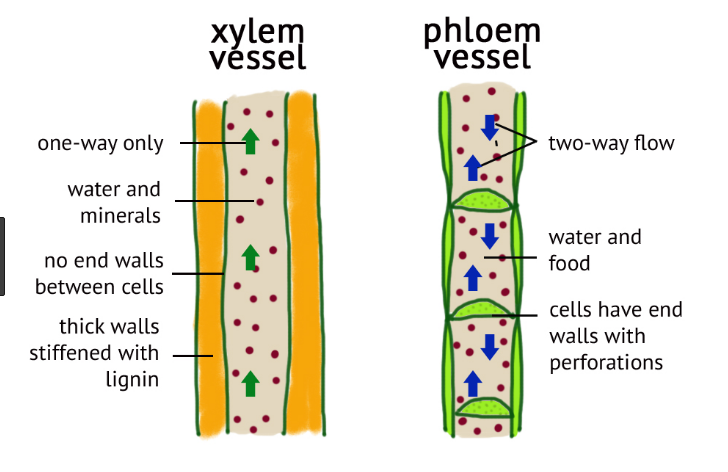
Xylem
Xylem is the principal water conducting tissue of the plant. It consists of four types of cells, viz..
(i) tracheids,
(ii) vessels,
(iii) fibres (xylem fibres), and
(iv) xylem or wood parenchyma
These constituents of xylem are also referred to as elements of xylem. The tracheids and vessels together are known as tracheary elements.
- Origin. The xylem which differentiates from the procambium of the apical meristem, is known as primary xylem. The secondary xylem develops from the vascular cambium after the completion of the primary growth.
- Structure. The detailed structure of various xylem elements is described below.
(a) Tracheary elements. The two basic types of tracheary elements are tracheids and vessel members.
Tracheids are characteristic of all vascular plants such as pteridophytes, gymnosperms and angiosperms. In primitive vascular plants xylemn is made only of tracheids (pteridophytes and gymnosperms). Tracheids originate trom single cells. These are elongated cells with tapering ends.The end walls are without perforations imperforate). Their length varies from 1 to 3 mm. Tracheids lack protoplast, and hence are dead.Fairly large cavity of these cells is without any contents. The wall of tracheids is moderately thick usually lignified. After the deposition of
and secondary wall thickenings, the cell organelles degenerate. Secondary wall depositions are of following types:
(i) Annular. In this type thickening material1 is deposited in the form of rings .
(ii) Spiral. The thickening is deposited in the form of a single or more helices or spirals.
The rings or helices may be arranged in a loose or compact manner. Annular and spiral thickenings are present in the first formed tracheary elements, i.e., protoxylem.
(iii) Scalariform. In later formed elements of xylem (metaxylem) the helical bands become joined in certain areas, giving the appearance of a ladder. This is known as scalariform thickening
(iv) Reticulate. In still later formed tracheary elements, the secondary wall thickenings are in the form of a network formed by the intercrossing of spirals.
(v) Pitted. In the late primary xylem and i the secondary xylem, the entire wall surface is thickened except for uniformly distributed thin areas known as pits. The density and arrangement of pits may vary in different walls of the same cell. The pits are of two types simple and bordered. The simple pits are the areas with only primary wall without any secondary thickening. The bordered pits are, however, more complicated as the secondary wall partially outgrows the pit to form a dome-shaped structure with a tiny perforation in the middle. The primary wall present between a pit pair develops a thickening in central part which is known as torus.
Physiologically, torus is very important as it acts as a valve membrane.
Vessels are long tube-like structures ideally suited for the conduction of water and solutes.These are made of a row of cylindrical cells formed arranged in longitudinal series. The partition walls these cells are perforated and as sucu
of tube-like.become the entire structure becomes as perforated region of the wall is known of pertoration plate. If the perforation plate contals kening gie large pore, it is called simple perforation plate . Sometimes perforation plate has many perforations and such a plate is known as multiple perforation plate . In different plants the perforation plate may be horizontal or oblique. Vessels with transvere end walls and simple perforations are considered to be more advanced than vessels with oblique end walls and multiple perforations.Vessels are found in the wood of almost all the angiosperms except certain primitive members of the order Ranales (vesselless dicots), Drimys, e.g Trochodendron, Tetracentron, Pseudowintera, etc. Vessels also occur in some pteridophytes, Such as Selaginella (S. rupestris, S. densa, S. arizonica,.rupicola), Equisetum, Pteridium aquilinum, Nephrodium, filix-mas (roots), Dryopteris olypodium, etc., and in the members of order netales of gymnosperms (e.g., Gnetum, Ephedra and Welwitschia).
Vessels are much shorter and broader than types of tracheids. Like tracheids various secondary wall thickenings (viz., annular, spiral,scalariform, reticulate and pitted) are also occur on the vessel walls.
(b) Fibres. Fibres are long cells with lignified secondary walls. Xylem fibres are of two types:
(i) fibre tracheids and
(ii) libriform fibres.Libriform fibres are longer and have thicker walls than the fibre tracheids. Fibre-tracheids possess bordered pits while libriform fibres have slit-like simple pits. Fibre tracheids provide mechanical strength while libriform fibres may conduct organic food.
(C) Parenchyma cells. Xylem parenchyma is represented by axial (or wood) parenchyma and ray parenchyma. The axial parenchyma cells are arranged in vertical series where as ray parenchyma are in radial transverse series. Both the types of cells are basically similar in structure and contents.These cells store starch, oils and many other
ergastic substances. The walls are lignified and may show Simple or bordered pits. Some of the parenchymatous cells become sclerified forming sclereids or sclerotic cells.Protoxylem and Metaxylem On the basis of size and development, xylem elements can be differentiated into protoxylem and metaxylem.
The first formed elements of primary xylem constitute the protoxylem; they develop directly from the procambium. In these elements of xylem lignification starts before the
completion of elongation, therefore,they are capable of being stretched. The tracheary elements i.e., tracheids and vessels) of primary xylem are relatively short and usually possess annular or roots), spiral thickenings. The metaxylem is later formed primary xylem. It develops after protoxylem.elements have been formed. It consists of longer and broader tracheary elements lignification occurs after completion of clongation. Hence these elements do not undergo further stretching. Tracheids and vessels constituting metaxylem have reticulate, spiral or pitted thickenings.
Phloem
Phloem is mainly responsible for the translocation of organic food synthesized by the plant. Together with xylem, it forms the vascular tissue. Like xylem, phloem is also composed of a variety of cell types. The constituent cells of the phioem are sieve elements (sieve cells, sieve tubes)companion cells, phloem fibres and phloem
parenchyma cells. On the basis of development, phloem is
grouped into two types primary phloem and secondary phloem. Primary phloem is produced by the procambium while the secondary phlolem develops from the vascular cambium.
- Structure. The detailed structure of various phloem elements is described below.
(a) Sieve elements. Sieve elements include sieve tube members and sieve cells. Both of these are specialized thin walled cells which have characteristic sieve areas in their walls.
Sieve tube members are long, slender, tube-like cells joined end to end to form long tubular channelsthe sieve tubes. Sieve tube members possess specialized sieve areas on the end walls called sieve plate. Sieve plate is known as simple if it has only a single sieve area and compound when the sieve plate has many sieve areas.
Young sieve tube members have abundant cytoplasm but their is no nucleus. The nucleus disintegrates during their development. members Adjacent sieve tube are interconnected by strands of cytoplasm through the pores of sieve plates. At maturity the sieve tubes have a large central vacuole and a thin layer of cytoplasm. Sieve tubes also possess some slime bodies.
During autumn season, callose (a polymer of cellulose) is deposited on and around the sieve areas, especially in temperate plants. lt ultimately forms a well defined plug or pad on the sieve plate. Formation of plug marks the end of the conducting activity of the sieve tube. Sieve cells are somewhat elongated and narrow With tapering ends. In these cells sieve areas occur all over the wall and, hence, sieve plate as such can not be distinguished. In pteridophytes and gymnosperms sieve cells remain separate but in angiosperms they form long sieve tubes.
(b) Companion cells. All angiosperms have specialized parenchyma cells, called companion cells, associated with the sieve tube elements. Usually a single companion cell extends through sieve tube the of the whole length but sometimes there is a vertical file of two or more companion cells next to each SIeve tube. These are living cells. The granular cytoplasm of these cells is continuous with that of the associated sieve tube through numerous The companion cells Small plasmodesmata. possess nucleus even at maturity. it has besides serving its own cytoplasm, also serves for the toplasr of the associated sieve tube Suggested that nucleus of the companion cell, (the latter lacks nucleus at maturity).strictly comparable to companion cells are not pteridophytes and gymnosperms, cells are not found. In conifers, however, a few cells similar to companion cells occur with sieve cells and are called albuminous cells.
(c) Phloem fibres. These are also known as bast fibres and occur in both the primary and secondary phloem. In primary phloem, fibres are generally present in outer part while in the secondary phloem, fibres are distributed in many different ways. Fibres are invariably long and the walls are thick. Phloem fibres of Linum,Cannabis, Hibiscus, etc., are of great commercial importance. Sclereids of different shapes and sizes may also be present mostly in the older parts of the phloem. These are formed due to lignification of parenchymatous cells
(d) Parenchyma cells. Parenchymatous cells containing various ergastic substances such as starch, tannins, crystals, etc., are very common in the region of phloem. These cells are commonly associated with fibres and sclereids and have lignified walls with secondary thickening In monocotyledons phloem parenchyma is absent.
Protophloem and Metaphloem
Just like primary xylem, primary phloem is also of two types protophloem and metaphloem.Protophloem elements mature early even when the plant part is undergoing elongation. Thus, sieve elements also get stretched and become nonfunctional. Finally, the protophloem elements get completely disfigured. Sieve elements of protophloem are narrow and indistinct. They may or may not be associated with companion cells. Metaphloem elements are generally wider and companion cells are regularly present.
FAQ.
- what are the 3 types of plant tissues and what are their main functions
- examples of plant tissues and their functions
- function of plant tissue
- What are the main functions of the three plant tissue?
- What is tissue and function?
- Which plant tissue has the most important function Why?
- What is the structure and function of plant cells and tissues?
- function of plant tissue culture
- functions of plant tissue xylem
- function of xylem and phloem
- Function of phloem
- what is Protophloem and Metaphloem
- what is Characteristics of collenchyma?
- what is permanent tissue?